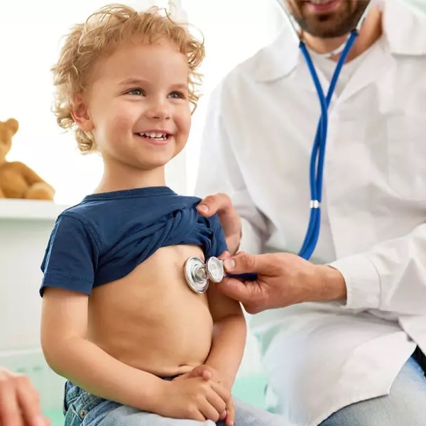
Device Closure of Heart Holes
Let your child breathe freely and live a healthy life after successful device closure treatment.
Atria or the upper heart chambers have a wall for separation called the atrial septum. Ventricles or lower heart chambers have two sides right and left, both separated by a membrane called the ventricular septum. The left atrium receives oxygenated blood from the pulmonary veins and supplies this blood to the left ventricle via the mitral valve to ensure proper circulation of the oxygenated blood throughout the body.
Even though the heart walls are properly separated to ensure a good circulation system, there are some situations of congenital heart defect or holes in the atrial or ventricle wall of the heart that can lead to disrupted flow of circulation. This disturbed blood circulation may also lead to heart failure and requires on-time treatment.
Small holes can be closed surgically with the process of device closure. The procedure involves the placement of a metal device in the location of the hole to restore the functioning of the heart.
About device closure of heart holes
During the surgery
- At the start of the process, you will be administered intravenous medicines and IV.
- Electrodes will be attached to your chest for ECG.
- A thin plastic tube called a catheter will be inserted through your groin/arm area.
- You will be on local anesthesia and sedatives to numb the operating area. However, you will be conscious throughout the surgical process.
- To get a clear picture of the heart chambers and the position and size of the hole, the cardiologist will inject a contrast dye into your arteries.
- If essential, a transoesophageal echocardiogram or echocardiogram is also performed.
- As soon as the size of the hole is known, a unique catheter is inserted into the arteries to transfer a small metallic mesh-like device, which is the closing device.
- The closing device is fixed in the hole and released with help of the catheter.
- The cardiac surgeon takes out the catheter. Eventually, heart tissue forms around the closure device and it becomes a part of your heart.

Note: There are numerous other symptosm that patients complain of such as edema, claudication, syncope, etc.
After the surgery

The opening through which the catheter is inserted is sutured once the tube is taken out.
You will be discharged after a couple of days from the surgery.
Listen to your doctor’s advice about going back to your routine post surgery.
During and after recovery, ensure you maintain a healthy lifestyle involving a good diet, exercising and regular heart checkups.
Types of device closure surgeries
Atrial Septal Defect (ASD)
In the case of ASD, there’s a small hole in the membrane dividing the atria – atrial septum. Due to this defect, the oxygenated blood starts leaking from the left atrium to the right atrium leading to reverse blood flow. Because of the leakage, the right atria gets overloaded with the responsibility of sending more blood to the lungs as deoxygenated and oxygenated blood is mixed up.
Symptoms of atrial septum blood:
- Heart murmurs
- Shortness of breath
- Easy fatigue after exercise
- Heart palpitations
Your doctor will prescribe ASD device closure before the symptoms worsen and irreversible.
There are four types of ASD, such as:
- Primum ASD: The problem starts in the lower side of the atrial septum and can get accompanied by some congenital heart disease.
- Secundrum ASD: In this condition, there’s a hole in the middle of the atrial septum. This one is the most commonly diagnosed ASD.
- Sinus Venous ASD: It is a rare defect that occurs in the upper region of the atrial septum – also accompanied by congenital heart issues.
- Coronary Sinus ASD: Another rare type of ASD in which the left atrium and the wall between the coronary sinus are absent. The coronary sinus is a part of your heart’s vein system.
Ventricular Septum Defect (VSD)
In the case of VSD, there’s a hole in the ventricular septum that mixes up oxygenated blood and deoxygenated blood leading to reverse blood flow from left to right. This further results in your heart pumping excessive blood to the lungs for purification which overloads the heart to supply more oxygenated blood across the body.
The defective hole in the ventricular septum can differ in size and in some rare cases, the wall between the ventricles is completely absent. It means there’s a single ventricle with both oxygenated and deoxygenated blood.
Symptoms of ventricular septum defect:
- Easy tiring
- Heart murmurs
- Poor eating
- Shortness of breath
- Not gaining weight easily
In case of a small hole in the ventricular septum, it can be fixed using device closure through heart catheterization.
Patent Ductus Arteriosus
When a baby is in the mother’s womb, a small duct, called ductus arteriosus, connects the pulmonary artery and aorta. The lungs of a baby start functioning after its born. Pure blood supply to the baby is from the mother through the placental connection.
When a baby is born, the ductus arteriosus closes in a couple of days or in some cases, within a year. If the opening doesn’t close on its own within a year, the pulmonary artery and aorta still have an open connection. The means the oxygenated blood in the aorta is leaked into the pulmonary artery, overloading the lungs to purify excess blood.
The main cause of PDA is still unknown, however, genetics is held responsible to some extent. The defect can be treated by device closure surgery.
The common symptoms noticed in a baby with PDA are:
- Fast breathing
- Bounding pulse
- Shortness of breath
- Not feeding well
- Sweating while feeding
Your cardiologist will diagnose PDA with EKG and Chest X-rays of the baby. For exact results, a contrast dye may also be administered through catheterization.
Coarctation
Coarctoplasty involves the insertion of a balloon-like tool on an end of the catheter and pushed into the vein. The main cause of coarctation is congenital. Initially, a child may not show any symptoms.
At a later stage, children have symptoms such as:
- Breathing difficulties
- Failure to thrive
- Chest pain
- Fainting episodes
- Dizziness
- Abnormal fatigue
- Headaches
- Nosebleeds
- Cold legs and feet
- Leg pain after exercise
If you or your child experience any of the above-mentioned issues, book an appointment with Dr Jagadesh and get a complete diagnostic evaluation followed by the best device closure options for treatment.
Let’s start a conversation…
We understand the importance of listening.
Feel free to contact us
